












Why They Cause Trouble at All
Wisdom teeth are the last to erupt, usually in the late teens or early twenties, into a jaw that has already finished growing. Whether there is room for them depends on jaw size, and frequently there is not.
Where there is no room, the tooth becomes impacted — blocked from erupting fully. It may angle forward against the molar in front, sit horizontally, remain entirely within the bone, or erupt partially with gum still covering part of the crown.
That last situation is the one that causes most problems. A partially erupted tooth has a flap of gum over it that traps food and bacteria and cannot be cleaned. Recurrent infection under that flap is the most common reason wisdom teeth are removed.
Fully impacted teeth sitting entirely within bone are a different matter. With no communication to the mouth, there is nothing to infect and nothing to clean. Many of these cause no problem at any point.
So the position matters more than the presence, and the position is visible on a radiograph. That is the whole basis of a sensible decision here.
Recurrent infection around a partially erupted tooth is also a common source of persistent bad taste and odour, because the flap traps debris that cannot be brushed out — one of the localised causes that resolves completely once the tooth is dealt with.
When Removal Is Clearly Indicated
These are situations where the tooth is causing damage or disease, and the question is when rather than whether.
Recurrent pericoronitis — infection under the gum flap of a partially erupted tooth. It recurs because the anatomy that causes it does not change, and each episode is painful and sometimes spreading. Two or more episodes is a clear indication.
Decay in the wisdom tooth or, more importantly, in the molar in front of it. A forward-angled wisdom tooth creates an uncleanable trap against the second molar, and decay there frequently damages a tooth that matters far more than the wisdom tooth does.
Cysts or other pathology around the crown of an unerupted tooth. These are uncommon, they are painless, and they are found on radiographs — which is one of the arguments for periodic imaging even where teeth are symptomless.
Resorption of the root of the second molar caused by pressure from the wisdom tooth. It is silent, it is progressive, and by the time it is advanced the more important tooth may be lost — which is why the assessment of the neighbouring tooth is part of this decision.
Where the second molar has already decayed against the wisdom tooth, both teeth are part of the plan. Removing the wisdom tooth gives access to restore the neighbour, and what that restoration involves depends on how much structure the decay has taken.
When Removal Is Not Indicated
The other side, and the one that changed in professional guidance. Removing a tooth that will never cause a problem exposes the patient to surgical risk for no clinical gain.
A fully impacted, symptomless tooth entirely within bone, with no cyst, no resorption of the neighbour and no communication with the mouth, is generally left alone and monitored radiographically.
A fully erupted wisdom tooth in a normal position that can be cleaned is a tooth. It functions, it can be restored if it decays, and it occasionally becomes useful later as a bridge abutment. There is no reason to remove it.
The old argument that wisdom teeth cause crowding of the front teeth has not held up. Late lower incisor crowding happens in people with and without wisdom teeth, and removing them prophylactically does not prevent it.
Where the decision is genuinely marginal, age is a legitimate factor: removal is easier and healing faster in younger patients, roots are less developed, and nerve proximity is often less. That argues for deciding rather than deferring indefinitely.
Monitoring is a real plan rather than an absence of one. A symptomless impacted tooth is reviewed on radiographs at intervals, which is how cysts and root resorption are found while they are still small — and that imaging is part of any full assessment regardless of why you came.
Assessing a Wisdom Tooth Before Removing It
A panoramic radiograph is the minimum, and it answers most of the questions: how the tooth is angled, how developed the roots are, whether there is decay or pathology, and how close the roots lie to the nerve canal in the lower jaw.
Where that radiograph suggests the roots are close to or overlying the inferior alveolar nerve, a CBCT is taken — a dental cone beam scan, at a small fraction of the dose of a medical CT. It shows the relationship in three dimensions rather than as a superimposition, and it changes the surgical approach where the relationship is intimate.
In the upper jaw the equivalent question is the sinus. Upper wisdom tooth roots can sit close to or within the sinus floor, and knowing that in advance changes how the tooth is removed and what the aftercare instructions are.
The second molar is assessed as carefully as the wisdom tooth. Decay on its back surface, root resorption and bone loss behind it all affect the decision and sometimes turn out to be the more important finding.
Symptoms are recorded but they do not decide it on their own. Pain in that region has several possible sources, and attributing it to a wisdom tooth without radiographic support is how symptomless teeth end up removed for nothing.
Where implants are also planned, the two assessments overlap. A wisdom tooth adjacent to a planned implant site affects the surgical sequence, and the implant plan is built after the extraction site has healed rather than around a tooth that is about to be removed.
What the Surgery Involves
It ranges from a routine extraction to a proper surgical procedure, depending entirely on the position, and the difference should be explained before rather than discovered.
| Situation | What is involved | Typical recovery |
|---|---|---|
| Fully erupted, normal position | Straightforward extraction, no incision | Like any extraction — days |
| Partially erupted, angled forward | Gum incision, sometimes sectioning the tooth | Swelling peaking at day 2–3 |
| Fully impacted in bone | Flap raised, bone removed, tooth sectioned | More swelling, up to a week of restriction |
| Horizontal impaction | Most involved — sectioning and staged removal | Similar, occasionally longer |
| Close to the nerve | CT planning; sometimes deliberate partial removal | As above, with specific monitoring |
| Upper, close to sinus | Careful technique; sinus precautions afterwards | Days, with restrictions on nose-blowing and flying |
Sectioning the tooth sounds worse than it is and is usually gentler. Removing a tooth in pieces along its own path requires far less force than removing it whole, and it protects the surrounding bone.
The Risks, Stated Honestly
Wisdom tooth surgery has genuine risks and they are worth knowing rather than glossing. They are also, for most teeth in most patients, low.
Nerve disturbance is the significant one in the lower jaw. The inferior alveolar nerve supplies sensation to the lower lip and chin, and the lingual nerve supplies the tongue. Temporary altered sensation happens in a small minority of cases; permanent change is considerably rarer.
The risk is not uniform — it depends on how close the roots lie to the canal, which is precisely what the CT establishes. Where the relationship is intimate, a coronectomy is sometimes proposed: the crown is removed and the roots deliberately left, avoiding the nerve entirely.
Dry socket is the more common complication, particularly for lower surgical removals, and it is reducible in large part, though it still occurs after some difficult lower removals with everything done correctly, and it is treated easily when it does. Infection is uncommon and treatable. Jaw stiffness for several days is normal rather than a complication.
In the upper jaw the specific risk is communication with the sinus, which usually closes on its own with sinus precautions. The anatomy involved is the same one that matters for upper implant planning.
Where an upper wisdom tooth sits within the sinus floor, its removal occasionally leaves a small communication that closes on its own with precautions. It matters more if grafting or implants are planned in that region later, which is another reason the assessment looks beyond the tooth itself.
Recovery, Realistically
More demanding than an ordinary extraction and entirely predictable in shape. Knowing the curve prevents most of the worry.
Swelling builds through the first day, peaks at day two or three, then falls. Jaw stiffness accompanies it and can make opening the mouth difficult for several days, which is normal and resolves. Bruising on the cheek or jaw appears in some patients.
Pain is managed with anti-inflammatory medication taken before the anaesthetic wears off rather than after. It should decrease each day from about day two. Pain that reverses direction around day three or four is the dry socket pattern.
Soft food for the first days and no chewing on the surgical side. No smoking, no straws, and no forceful rinsing on the first day — the same rules as any extraction and they matter more here because dry socket risk is higher.
Most people take two or three days away from demanding activity. Full comfort returns over one to two weeks, and the detailed aftercare instructions cover the whole period including what to report.
Antibiotics are not routine and should not be. They are prescribed where there is genuine infection risk — an existing infection, extensive bone removal, or a medically-compromised patient — and where prescribed, the full course is taken even once you feel well.
Having It Done While Travelling
Wisdom tooth surgery is workable on a treatment trip but it needs planning around, and it interacts badly with a tightly packed schedule.
The main consideration is timing relative to your flight home. Swelling peaks at day two or three, so surgery on the last day of a trip means travelling at the worst point. Scheduling it early in a visit is considerably more comfortable.
Flying itself is generally fine after a day for lower teeth. Upper wisdom teeth close to the sinus are the exception — where a sinus communication has occurred, the interval before flying is longer and specific.
It also interacts with the rest of the plan. Surgery on one side means chewing on the other for several days, which complicates fitting restorations or assessing a bite in the same week.
Where it is being combined with other treatment, the sequence usually puts the surgery early and the restorative work later — how the days divide up sets out how the treatments fit together.
Coronectomy: Removing the Crown Only
Worth knowing about because it is not widely explained and it is the right answer in a specific and identifiable group of cases.
Where the roots of a lower wisdom tooth are intimately related to the nerve canal, removing them carries a meaningfully higher risk of permanent sensory change. Coronectomy removes the crown — the part causing the problem — and deliberately leaves the roots in place.
The roots are left below the bone level and bone grows over them. They are not diseased; the pathology was around the crown. Long-term follow-up shows the retained roots usually remain quiet.
Occasionally they migrate away from the nerve over subsequent years, and if they ever need removing later they can be taken out at a safer distance. A small proportion require that second procedure.
It is not appropriate where the tooth is infected around the roots, or where the tooth is mobile. Where it is appropriate, it trades a small chance of a second procedure for a substantially lower risk of permanent numbness, and most patients take that trade.
Sedation and Anxiety
Wisdom tooth surgery is the procedure patients most often ask about sedation for, and it is a reasonable request rather than an indulgence.
Local anaesthetic alone is sufficient for the surgery itself — the area is genuinely numb and the procedure is not painful. What sedation addresses is the experience: the sounds, the pressure, and the anxiety of a longer surgical appointment.
It changes what you need to arrange. Fasting beforehand, someone to accompany you afterwards, no driving, and a day without commitments. For a patient travelling alone that last requirement needs thinking about in advance.
It also makes longer or bilateral procedures more comfortable, which can mean removing more than one tooth in a single appointment rather than across several.
Whether it is appropriate depends on your medical history as much as your anxiety, and that assessment happens at the consultation rather than on the day.
Nitrous oxide sits between local anaesthetic alone and intravenous sedation. It wears off within minutes, needs no fasting and no escort, and takes the edge off a long appointment — which makes it the more practical option for someone travelling without a companion.
Removing One or All Four
A question with no single right answer, and it depends on the position of each tooth rather than on convenience.
Where all four are impacted and all four are causing or will cause problems, removing them together — often under sedation — means one recovery period rather than several. That is a genuine argument, particularly for a patient travelling.
Where only one is problematic, removing the others because they happen to be there is prophylactic removal by another name, and the same reasoning applies as elsewhere on this page.
Removing both teeth on one side rather than both uppers or both lowers has a practical advantage: you can chew on the other side throughout. Doing both sides at once removes that option for several days.
Uppers are generally simpler and heal faster than lowers. Where a plan involves all four, doing the more difficult lower teeth with the corresponding uppers on the same side is usually the most comfortable arrangement.
For a visiting patient the calculation shifts towards doing them together. One recovery period inside one trip is easier to plan around than two visits, and the trip schedule is usually built that way where all four genuinely need removing.
Wisdom Teeth on a Treatment Trip
Wisdom teeth are a common addition to a Turkish treatment plan and one that deserves a specific caution: a wisdom tooth that is causing no symptoms and shows no pathology on the radiograph does not need removing because you happen to be in İstanbul. Prophylactic removal of asymptomatic, disease-free third molars is not supported, and a plan that includes all four without a reason for each has added them because you were here.
Where there is a reason — recurrent pericoronitis, decay in the wisdom tooth or the molar in front of it, a cyst, or a tooth that is going to be in the way of planned work — the case for doing it during the trip is strong, because the surgical time and the follow-up are together rather than months apart. A CBCT is taken first where the roots sit near the inferior alveolar nerve, and the scan is discussed with you rather than filed.
Timing against the flight matters more here than for almost anything else. Swelling and trismus peak on the second or third day, so surgical removals are scheduled early in the trip. Flying itself is not a problem for the site, but a long journey on the worst day is a miserable way to spend it, and there is no clinical reason to arrange it that way. The aftercare that follows is the same wherever you are.
Dry socket is the complication people ask about most. It is uncommon, most of the risk is reducible, and it is treated easily — but it typically appears on day three or four, which for many patients is after they have flown home. The out-of-hours number covers that, and the dressing it needs is something any dentist can place.
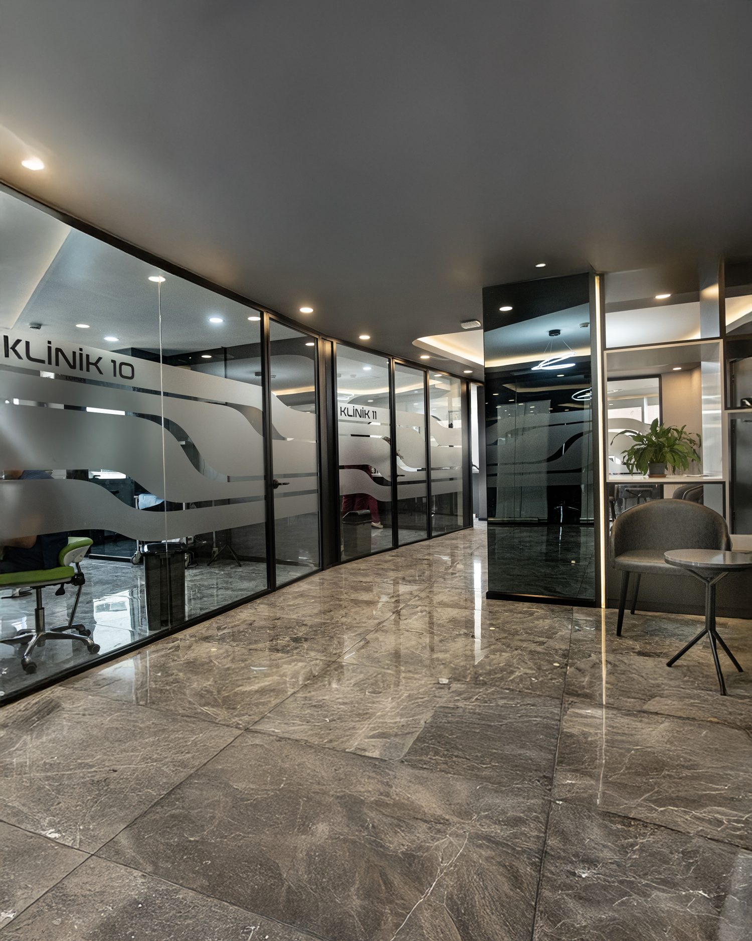
How We Approach Wisdom Teeth at Bağcılar
From the radiograph, tooth by tooth, with the second molar assessed as carefully as the wisdom tooth itself. Where a tooth is symptomless, fully impacted and causing no damage, we say so rather than proposing removal.
Where the panoramic radiograph suggests the roots lie close to the nerve canal, a CBCT is taken — a dental cone beam scan, at a small fraction of the dose of a medical CT before any decision. That relationship changes both the risk and the technique, and it cannot be judged reliably from a two-dimensional image.
Coronectomy is offered where it is appropriate rather than only where a patient asks about it, and the trade-off is explained: a small chance of a later procedure against a substantially lower risk of permanent sensory change.
For visiting patients, surgery is scheduled early in the trip rather than at the end, so that the swelling peak does not coincide with the flight home. Where it is combined with restorative work, the sequence accounts for the recovery.
Aftercare instructions are given in your own language, in writing, before you leave — and the records you take home include the radiographs and the operative note, which any dentist would need if anything required attention later.