










CBCT: The One That Changes Decisions
Cone beam computed tomography produces a three-dimensional image of the jaws. That sounds incremental against a conventional radiograph and it is not — it is the difference between a shadow and a measurement.
A panoramic radiograph flattens the jaw into a single plane. Bone width is invisible on it, the nerve canal is a superimposed line rather than a located structure, and the sinus floor is an approximation. All three are exactly what implant planning depends on.
A CBCT shows bone height, width and density at any point, the precise position of the inferior alveolar nerve, and the sinus anatomy in three dimensions. From that, the number of implants, their angles and whether grafting is needed can be determined rather than estimated.
It also changes wisdom tooth surgery. Where roots appear to overlie the nerve on a panoramic image, a CBCT establishes whether they genuinely do — which determines both the risk and whether a coronectomy is the better option.
We have it in the clinic. That matters practically as well as clinically: the scan happens at the assessment appointment rather than at a separate imaging centre on another day, which for a patient on a treatment trip removes a day from the schedule.
It changes bone assessment as well as nerve mapping. Whether grafting is needed, and how much, is a measurement rather than an impression once the scan exists — which is why a quote produced before one is a figure rather than a plan.
What a CBCT Does Not Do
Worth stating, because it is sometimes presented as answering everything and it does not.
It does not show soft tissue in useful detail. Gum thickness, attachment levels and pocket depths are measured with a probe, not read from a scan, and that charting remains the basis of any periodontal assessment.
It does not diagnose decay reliably. Small carious lesions between teeth are better seen on conventional bitewing radiographs, which have higher resolution for that specific job at a lower radiation dose.
It carries a higher dose than a panoramic radiograph, which is why it is taken where it changes decisions rather than routinely. Implant planning, complex surgery and specific diagnostic questions justify it; a general check-up does not.
And it does not plan anything by itself. The scan is data; the plan comes from someone reading it against your bite, your gum health and what you actually want — which is why the consultation matters more than the machine.
Intraoral Scanning
A small camera passed over the teeth builds a digital model of both arches in a few minutes. It replaces the tray of impression material that most people remember unfondly.
The patient-facing benefit is obvious: nothing to gag on, nothing setting in your mouth, and it can be paused and resumed. For anyone with a strong gag reflex it converts an ordeal into a non-event.
The clinical benefit is less visible and more significant. Conventional impressions distort slightly — the material shrinks, the tray flexes, the cast is poured from it and introduces its own error. A digital scan removes that chain.
It also makes the model reviewable immediately. A margin that was not captured cleanly shows on the screen while you are still in the chair, rather than being discovered by the technician the following day.
And it feeds directly into design. The same scan is used for restorative planning, for aligner treatment and for the digital design stage of a smile design, without re-recording anything.
The Laboratory in the Building
This changes the process more than any single machine, and its value is easiest to describe in terms of what goes wrong without it.
Shade is the clearest case. A restoration that comes back not quite matching has to go somewhere to be corrected. Where the laboratory is external, that means a courier cycle and another appointment. Where the technician is in the building, it means an hour.
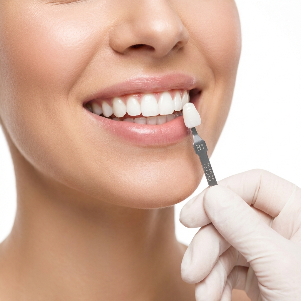
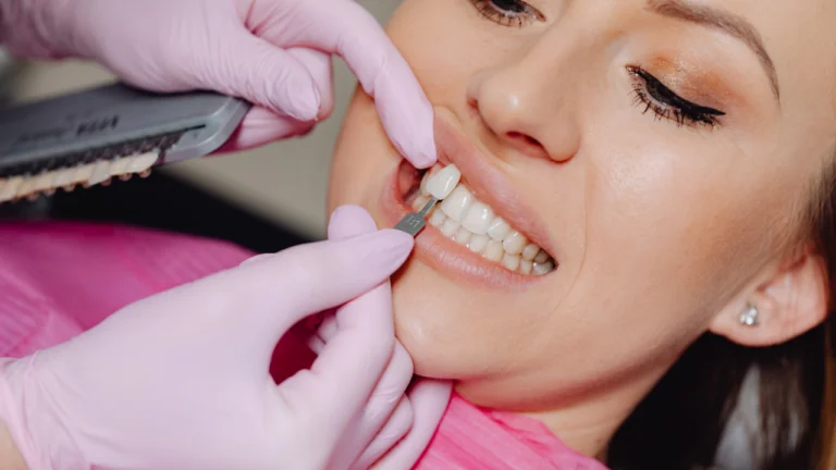
The technician can also see you directly. Shade taken by a technician looking at your teeth in daylight, rather than from a written shade code, produces a different quality of match — particularly on front teeth where the gradient from neck to edge matters.
It compresses the timetable, which for a patient travelling is the practical point. Preparation, laboratory work, try-in and fitting inside one trip is only realistic when the laboratory is not adding transit time between each stage.
What it does not do is guarantee quality. An in-house laboratory with a mediocre technician produces mediocre work faster. The advantage is the feedback loop, not the postcode — the material and how it is made still determine the result.
What Each Piece Actually Changes
The table separates what affects the clinical outcome from what affects the experience or the schedule. Both are worth having; they are not the same claim.
| Equipment | What it changes | Outcome or experience? |
|---|---|---|
| CBCT / 3D scan | Makes implant and surgical planning measurable rather than estimated | Outcome — significantly |
| Panoramic radiograph | Overview of all teeth, roots, bone levels and sinuses in one image | Outcome — the baseline for any full assessment |
| Intraoral scanner | Removes impression distortion; model reviewable immediately | Both |
| In-house laboratory | Same-day corrections, technician sees the patient, shorter trip | Both |
| Digital design software | Lets the proposal be discussed before anything is prepared | Experience, and it improves communication |
| Sedation facilities | Makes long or difficult appointments tolerable | Experience — the dentistry is unchanged |
| Surgical suite | Sterile field for implant surgery, separate from treatment chairs | Outcome |
A clinic listing only experience-level equipment while lacking a CBCT is telling you something about its priorities, and the gap is easy to check by asking one question.
Sterilisation Equipment
The least photogenic part of any clinic and the one people worry about most. The equipment matters less than the verification, which is the part rarely described.
An autoclave sterilises under pressurised steam at 134°C. Every licensed clinic has one; the question is not whether it exists but whether every cycle is confirmed to have worked.
The indicator on each pouch confirms that the pack has been through a cycle, which is why pouches are opened in front of you; what confirms the cycle reached temperature, pressure and hold time is the autoclave's own cycle record together with an indicator placed inside the load. That is a per-load check and it is visible to you — pouches should be opened in front of you with the indicator showing.
Biological indicators are the more searching test: spores that are cultured afterwards to confirm the autoclave actually kills them. Run periodically, logged, and available if you ask.
Everything disposable is single-use — needles, gloves, suction tips, prophylaxis cups, surgical drapes. The full protocol is set out separately, step by step, because it is one of the few things a patient can genuinely verify.
The Surgical Suite
Implant surgery is surgery, and the setting for it differs from a general treatment chair in ways that matter.
A sterile field is established — sterile drapes, sterile instruments opened at the chair, gowns and a scrub procedure. The point is that the implant site is isolated from everything that has not been sterilised.
The room is separate from routine treatment, which reduces airborne contamination and allows the surgical setup to be prepared without competing with a check-up schedule.
Surgical instrumentation is specific: the implant motor with controlled torque and speed, and the drill sequence for the system being used. Placing an implant with uncontrolled speed generates heat, and overheated bone does not integrate.
It applies equally to grafting and to sinus procedures, where the membrane work requires both the field and the instrumentation to be right.
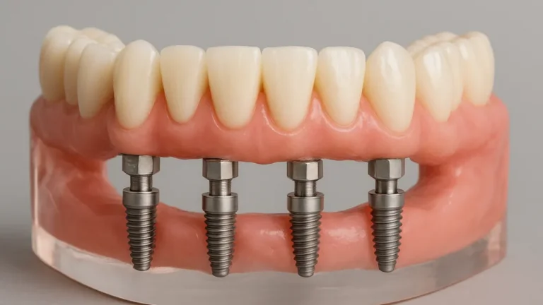
Where a full arch is being placed, the setup matters more again. Full-arch surgery involves extractions, several implants and a temporary bridge in one session, and the field has to hold for the whole of it.
Digital Design and What It Is Good For
Design software overlays a proposed tooth arrangement onto photographs of your face, so proportion can be judged against your actual features rather than against an abstract ideal.
Its real value is conversation. You see a rendering, say what you dislike, and the design changes — before anything irreversible. Disagreements about width, length or shade are far cheaper to resolve on a screen than in ceramic.
What it does not do is predict the outcome. A render is a two-dimensional image of a three-dimensional result that also has to function, and software does not know how your lip moves or how your teeth meet.
Which is why the digital stage is followed by a physical one. The design is transferred to a wax model and then into your mouth as a trial smile you wear for an hour — that step is the one that matters, and no software replaces it.
Treat an impressive simulation as a starting point rather than a promise. A clinic presenting a render as the guaranteed result has overstated what the tool does.
Equipment Claims Worth Checking
Equipment is one of the easier things to verify, and a few specific questions separate a real capability from a marketing line.
- Is the CBCT in the clinic, or taken at an external centre on another day?
- Is the laboratory in the building, and will the technician see me for shade?
- Will impressions be digital or conventional — and if conventional, why?
- Is implant surgery done in a separate surgical suite?
- What chemical and biological indicators are used, and how often is biological testing run?
- Which implant system will be used, and will I receive the implant passport?
The first is the one most often overstated. "We use CBCT" and "we have a CBCT" are different claims, and for a patient on a short trip the difference is a day.
The second matters for the schedule. A clinic quoting a one-week veneer plan while sending work to an external laboratory is describing a timetable with no room for a shade correction.
None of these require specialist knowledge to ask or to interpret. The wider set of checks covers what else is worth establishing before committing.
Two pieces of equipment on this list are worth asking about specifically, because they are where the marketing runs furthest ahead of the substance: the laser and the milling unit. Which laser wavelength does what, and where a blade is still the right instrument goes through the first in detail, and it is the page to read before accepting a quote with a laser line on it.
Where Equipment Makes No Difference
The honest counterweight to the rest of this page, and the part most clinic technology pages leave out.
Equipment does not decide whether a treatment was correctly indicated. A CBCT in the hands of someone planning twenty crowns for a patient who needed four produces a very well-imaged over-treatment.
It does not improve a preparation. Margin accuracy, how much enamel is removed and whether the reduction follows the final design are technique, and technique is a person rather than a machine.
It does not substitute for the assessment. Probing depths, bite analysis and asking what actually bothers you are the parts of the appointment that determine the plan, and none of them involve a device.
So read an equipment list as a floor rather than a ceiling. It tells you what is possible in the building. What is actually done with it shows up in the written treatment plan instead.
The same applies to material choice. No scanner decides whether a molar should be monolithic zirconia or a front tooth glass-ceramic; that decision comes from position, load and the state of the tooth underneath.
Radiation, Honestly
A reasonable question that is usually answered defensively. The straightforward version is more reassuring than the deflection.
Dental radiographs are low-dose. A panoramic image is a small fraction of the radiation you receive from the natural environment over a year, and a single bitewing is smaller still.
A CBCT is higher than either, though still substantially below a medical CT of the same region. That is precisely why it is taken where it changes a decision rather than as a routine screening image.
The field of view is limited to the area being planned rather than scanning the whole jaw where only one region is relevant. A smaller field means a lower dose and better resolution at the same time.
If you are pregnant or may be, say so before any imaging. Elective radiographs are deferred, and the assessment is planned around that — it is a routine conversation rather than an obstacle.
What We Have at Bağcılar
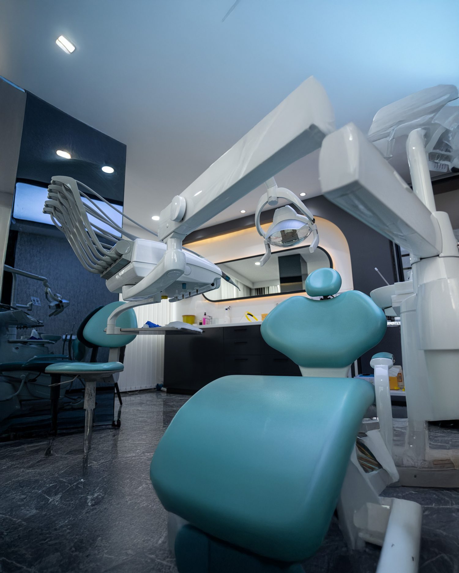
CBCT for three-dimensional imaging, in the clinic, so implant and surgical planning happens at the assessment appointment rather than on a separate day at an external centre.
A panoramic radiography unit for the overview image that begins any full assessment, and conventional radiographs where an individual tooth or the contacts between back teeth need detail.
An intraoral scanner, so impressions are digital. That removes the impression material entirely, removes a source of distortion, and lets the model be checked on screen before you leave the chair.
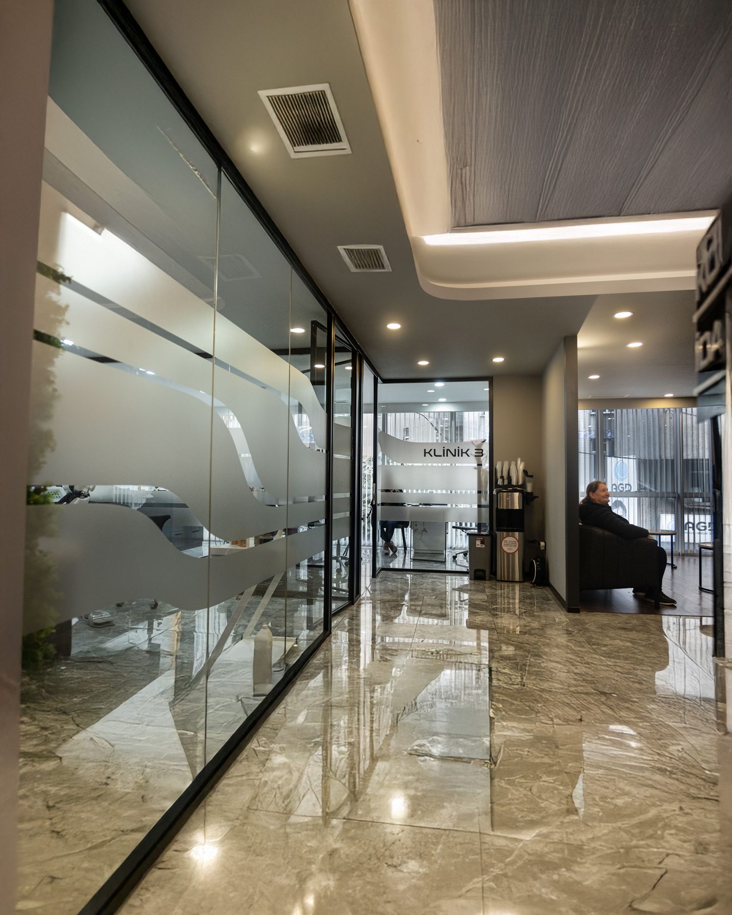
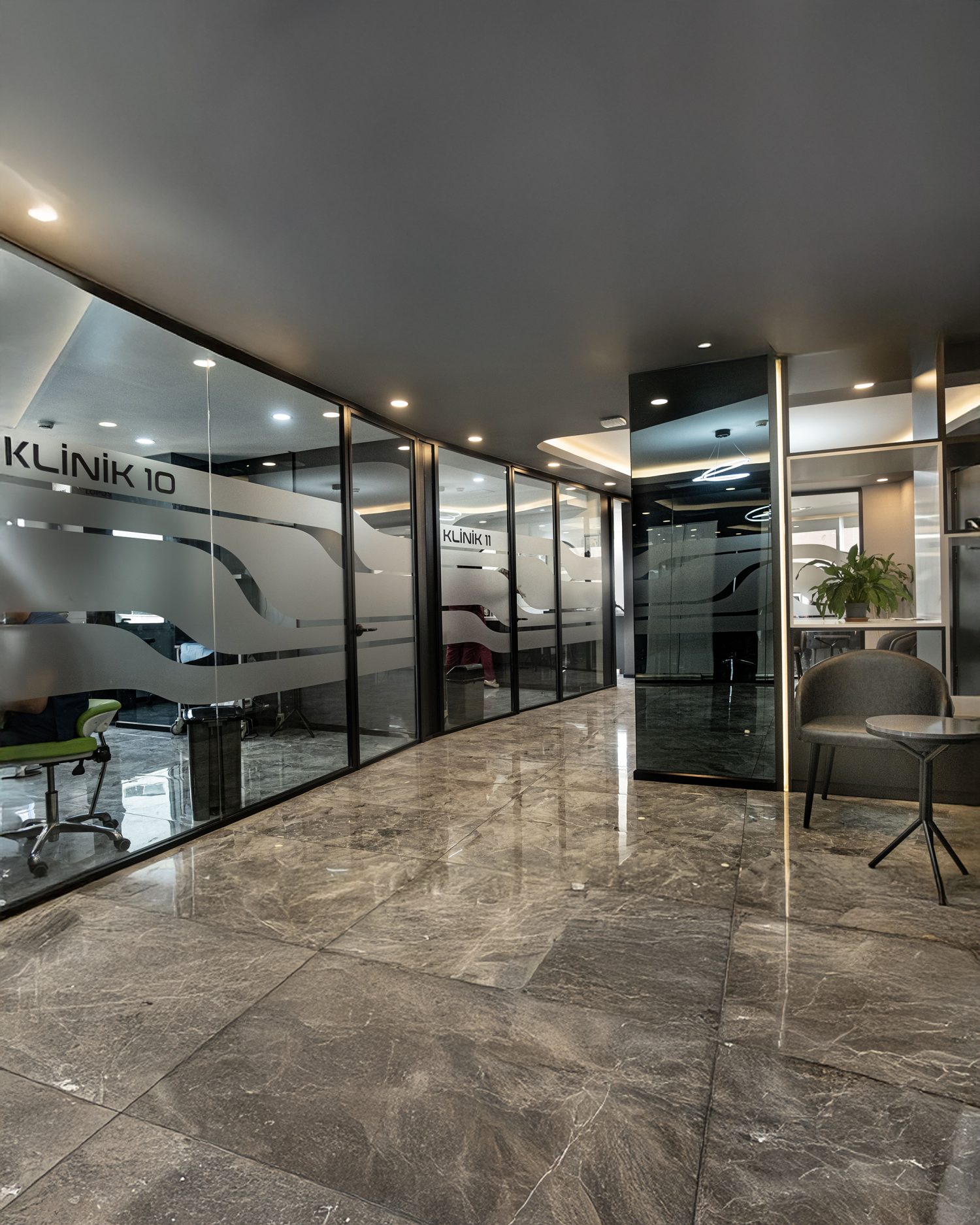
A CAD/CAM laboratory in the building, with the technician available to see you in person for shade. Corrections take hours rather than a courier cycle, which is what makes a single-trip restorative plan realistic.
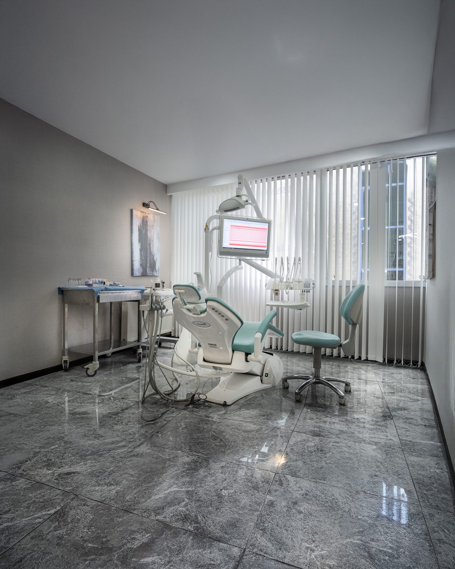
And a surgical setup for implant placement and grafting, separate from routine treatment. What is done with all of it is set out tooth by tooth in your written plan, and everything placed carries a lifetime guarantee on materials and workmanship — with the exclusions stated plainly.
What to Read Into an Equipment List
Equipment lists on clinic websites are written to reassure rather than to inform, and reading one usefully means asking a different question: what does this let them do that they could not do otherwise?
A CBCT answers that clearly — it makes implant planning measurable. An intraoral scanner answers it partly. A photograph of a treatment room with a modern chair answers it not at all, and most lists contain more of the third than the first.
The absence of certain items is the more informative signal. A clinic offering implant treatment without a CBCT on site is either sending patients elsewhere for imaging or planning without it, and both are worth knowing before you book.
Equally, the presence of everything means little on its own. What determines your result is the assessment and the plan, and neither of those shows up in a review or a photograph any more than it shows up in an equipment list.
The useful test is whether a clinic can explain what each thing changes for you specifically. That answer is short, concrete and impossible to fake — and it is the same test this page has tried to apply to itself.
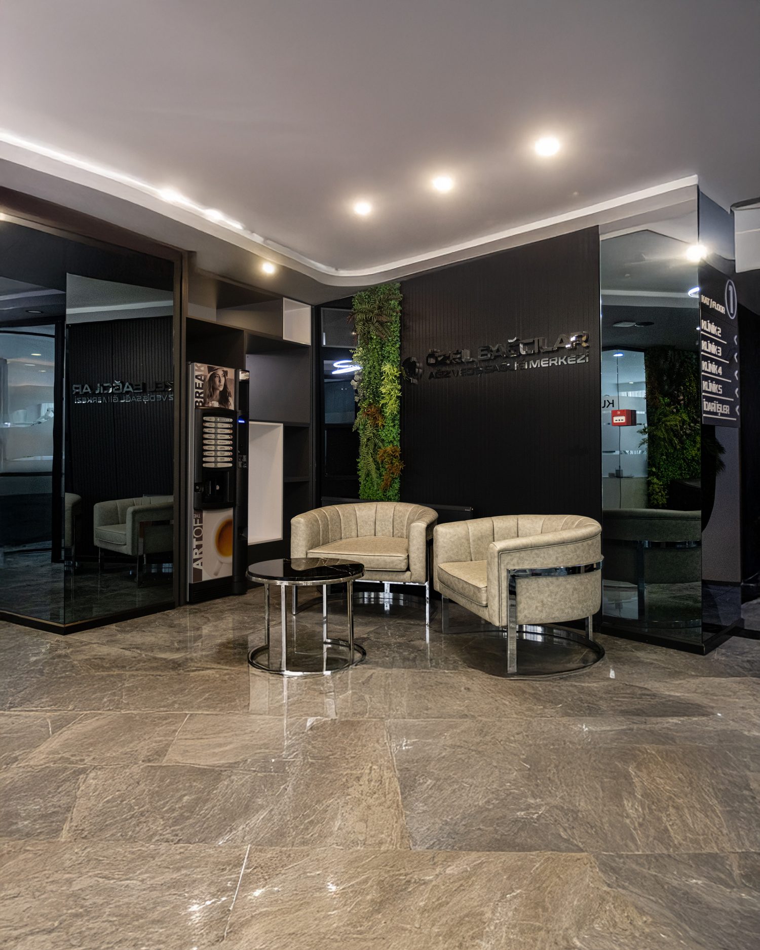
Equipment lists are the easiest thing in the world for a clinic website to write, and İstanbul has a great many of them. The useful test is not whether a machine is named but whether you encounter it. A CBCT that exists changes nothing; a CBCT whose images are on the screen while your plan is being explained changes the plan. An in-house laboratory is a floor of the building in Bağcılar with technicians on it, and you can be introduced to the person making your crown — which is a claim that either survives being asked about or does not. When you compare Turkish clinics, ask to be shown the thing rather than told about it.