











Why Bone Disappears
Bone is not a static scaffold. It is living tissue that constantly remodels, adding material where it is loaded and removing it where it is not. A tooth root transmits chewing force into the surrounding bone, and that stimulus is what tells the body to maintain it.
Remove the root and the stimulus stops. Within weeks, resorption begins. The bone that formed the socket wall — thin, and dependent on the tooth for its blood supply — is lost first, and the ridge narrows from the outer surface inwards.
The scale surprises people. Published measurements show substantial width loss in the first six months and continuing, slower loss over subsequent years. The front of the upper jaw loses the most, because the outer plate of bone there is thinnest to begin with.
This is the single strongest argument for not delaying a decision about replacement. Bone that is still present is far easier to work with than bone that has to be rebuilt, and the difference is measured in months of treatment and in predictability.
There is one exception worth knowing. A tooth removed because of a failed restoration or fracture, where the surrounding bone was healthy, leaves a much better site than one removed after years of infection. The bone was intact until the day of extraction, which is why these cases are frequently the best candidates for immediate placement.
Other Reasons Bone Is Lost
Extraction is the commonest cause but not the only one, and the cause affects how the defect is shaped — which in turn affects how easily it can be rebuilt.
- Periodontal disease. Destroys bone around teeth that are still present, often producing broad horizontal loss that is considerably harder to regenerate than a contained socket. Gum disease must be stabilised before any graft.
- Infection. A long-standing abscess destroys surrounding bone and leaves infected tissue that must be thoroughly removed before grafting.
- Trauma. A fractured jaw or an avulsed tooth can take the supporting plate with it.
- Denture pressure. A denture rests on the ridge and loads it in compression rather than through roots, which accelerates rather than prevents resorption.
- Failed implants. A failed implant leaves a defect the size of the implant plus whatever infection removed.
The shape matters enormously. A contained defect with walls on several sides regenerates predictably, because the graft material is held in place and blood vessels grow in from the surrounding bone. A broad flat ridge with no walls is the hardest scenario and the least predictable.
When Grafting Is Genuinely Needed
An implant needs bone around it in every direction, not just underneath. The usual requirement is at least a millimetre or two of bone on the outer and inner surfaces, and enough height to place an implant of adequate length without reaching the sinus above or the nerve below.
Where those minimums are not met, the choice is to graft, to place a shorter or narrower implant if the anatomy allows, or to place the implant somewhere else entirely. All three are legitimate and the decision is made on the 3D scan.
Being clear about the threshold matters because grafting is sometimes proposed where it is not needed. A patient with adequate bone who is told they need a graft is being sold an unnecessary procedure, and asking to see the measurement on the scan is entirely reasonable.
Equally, grafting is sometimes avoided where it is needed, by placing an implant into insufficient bone and hoping. That produces an implant with exposed threads and a poor long-term outlook, and it is the more damaging error of the two.
Where the tooth being replaced was lost to a previous implant failure, the assessment is different again — infection has usually removed more bone than the implant itself occupied, and the site needs thorough cleaning before any graft is placed.
The Materials Used
Graft material provides a scaffold. In most cases it does not become bone itself — it holds the space while your own bone grows into and through it, and is gradually replaced.
Autograft is your own bone, taken from elsewhere — usually the chin, the back of the lower jaw, or the hip in large cases. It contains living cells and is the biological gold standard, at the cost of a second surgical site and its own recovery.
Xenograft is processed animal bone, most commonly bovine. The organic component is removed entirely, leaving a mineral scaffold that resorbs very slowly and holds volume well. It is the most widely used material in dental grafting and has decades of clinical data behind it.
Allograft is processed human donor bone from tissue banks, and alloplast is fully synthetic — usually calcium phosphate ceramics. Both avoid a second surgical site and both perform well in appropriately selected cases.
Which material is chosen matters less than patients expect, provided it is one with an established record. Containment, tension-free closure and the absence of smoking influence the outcome far more than the choice between a bovine mineral and a synthetic ceramic. A clinic that markets a particular graft material as its distinguishing feature is emphasising the least variable part of the procedure.
Socket Preservation — The Easiest Case
The simplest and most effective grafting is done at the moment a tooth is removed, before any bone has been lost. It is called socket preservation, and it is prevention rather than reconstruction.
Graft material is placed into the empty socket immediately after extraction and covered with a membrane or soft tissue. It maintains the shape of the ridge while healing, so that when the implant is placed several months later the site is close to its original dimensions.
The difference in outcome is substantial. A preserved socket usually needs no further grafting; an unpreserved one frequently needs ridge augmentation, which is a larger procedure with a longer healing period and a less certain result.
This is why the conversation about replacement should happen before the extraction rather than afterwards. A patient who knows an implant is planned can have the socket preserved at no extra surgical visit. One who decides two years later is starting from a considerably worse position, and often requires additional procedures in the upper jaw as well.
Ridge Augmentation
Where bone has already been lost, rebuilding it is a larger undertaking and the technique depends on which dimension is missing.
Guided bone regeneration is the workhorse. Particulate graft material is placed against the deficient area and covered with a membrane that keeps soft tissue out while bone grows in. Soft tissue grows much faster than bone, and without a barrier it fills the space first — which is what the membrane prevents.
Block grafting uses a solid piece of bone screwed into place. It provides more structural support for large defects and holds its volume reliably, at the cost of a more involved procedure and, if autogenous, a donor site.
Ridge splitting is used where the ridge is adequate in height but too narrow. The ridge is separated along its length and expanded, with graft material placed in the gap created. It suits specific anatomy and avoids rebuilding from the outside.
Comparing the Approaches
| Socket preservation | Guided regeneration | Block graft | Ridge split | |
|---|---|---|---|---|
| When it is done | At extraction | Any time after loss | Larger defects | Narrow ridges |
| Second surgical site | No | No | Sometimes | No |
| Healing before implant | 3–4 months | 4–6 months | 6–9 months | 3–4 months |
| Predictability | High | Good | Good | Technique-sensitive |
| Implant at same time | No | Sometimes | Rarely | Often |
| Volume gained | Maintains existing | Moderate | Substantial | Width only |
The pattern is consistent: the earlier the intervention, the smaller the procedure and the shorter the healing. Every row rewards acting before bone is lost rather than rebuilding afterwards, which is the practical lesson of the whole page.
One column deliberately absent is cost, because we do not publish price lists and a single figure would mislead. What the table does show is the relationship: the procedures get larger and slower as you move right, and every one of them is avoided by not losing the bone in the first place.
The "implant at same time" row is the one that changes travel plans. Where the implant can be placed with the graft, treatment is two trips. Where it cannot, it is three — and that is worth establishing from a scan before flights are booked rather than discovering it at the first appointment.
What the Procedure Involves
Most dental grafting is done under local anaesthetic, with sedation available. A socket preservation adds only minutes to an extraction appointment. Larger augmentation takes an hour or more.
- The gum is lifted to expose the deficient bone.
- The site is cleaned of any granulation or infected tissue — the step that most affects success.
- Small perforations are sometimes made in the bone surface to encourage blood vessels to grow into the graft.
- Graft material is placed and shaped to the required contour.
- A membrane is placed over it and secured where necessary.
- The gum is closed over the site, ideally without tension.
That last point is more important than it sounds. A graft covered by tissue under tension tends to open during healing, which exposes the material and usually loses it. Achieving tension-free closure over an augmented ridge often requires releasing the tissue, and it is a significant part of the technique.
Healing and Timeline
Graft healing is a slow biological process and it cannot be accelerated by wanting it to be faster. Blood vessels grow into the graft, cells follow, and new bone forms progressively from the surrounding walls inwards.
Socket preservation is generally ready for an implant at three to four months. Guided regeneration takes four to six. Block grafts and large augmentations take six to nine, sometimes longer where the volume rebuilt is substantial.
The first two weeks are the critical period. Swelling peaks at forty-eight hours and settles over a week or so. The site must not be disturbed: no pressing with the tongue, no chewing on that side, no smoking, and any temporary denture must be adjusted so it does not rest on the graft.
Before the implant is placed, the site is reassessed on a new 3D scan rather than by the calendar. Grafts do not all heal at the same rate, and placing an implant into a graft that has not fully consolidated is a predictable route to failure.
Risks and What Can Go Wrong
Grafting is generally safe and predictable, but it has a specific set of complications and they should be understood before consenting.
- Membrane exposure. The commonest complication. The tissue opens over the graft, exposing the membrane to the mouth. Small exposures often heal; larger ones can lose part of the graft.
- Infection. Uncommon but serious, usually requiring removal of the graft material and a fresh start after healing.
- Incomplete regeneration. Less bone forms than planned. This may still allow implant placement, or may require a second graft.
- Donor site discomfort. Where autogenous bone is taken, that site has its own recovery — usually the more uncomfortable of the two.
- Graft failure. Rare in contained defects, more common in large reconstructions and markedly more common in smokers.
Smoking deserves emphasis because it affects grafting more than almost any other dental procedure. It restricts the blood supply the graft depends on entirely, and failure rates in smokers are substantially higher. Many clinics decline large grafts in continuing smokers, and that is a clinical judgement rather than a moral one.
Patients who grind heavily are also at raised risk, though for a different reason — the eventual implant sits in regenerated bone, which is initially less dense than native bone and less tolerant of excessive load. Protection is planned alongside the restoration rather than after.
When Grafting Can Be Avoided
Not every deficient site needs rebuilding, and the alternatives have improved considerably. Any thorough plan should have considered these before proposing a graft.
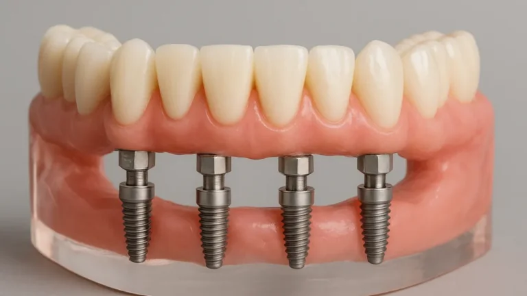
Angled implants. Placing implants at an angle to engage bone that is still present, rather than rebuilding bone that is not. This is the principle behind All-on-4 treatment and it avoids grafting in a large proportion of full-arch cases.
Short implants. Modern short implants perform well in situations that previously required vertical augmentation. They are not suitable everywhere, but where they are, they replace a six-month graft with nothing at all.
Zygomatic implants. In the severely resorbed upper jaw, anchoring in the cheekbone bypasses the jaw entirely. Reserved for severe cases, but genuinely avoids extensive grafting.
A different restoration. Sometimes the honest answer is that a bridge or a well-made denture serves the patient better than a year of staged surgery. That comparison deserves to be made explicitly rather than assumed away.
Where the site in question is an upper back tooth, a sinus lift is a specific form of grafting with its own decision points, and it is often combined with implant placement in a single procedure where enough bone remains.
Planning a Graft From Another Country
Grafting complicates international treatment because it inserts a long healing period into the middle of the plan, and it is the main reason a treatment described as "one week" becomes a year.
The usual structure is three trips rather than two: extraction and graft, then implant placement several months later, then the restoration after integration. Some of these can be combined depending on the case — an implant placed at the same time as a small graft, for instance — but the healing periods cannot be compressed.
This is worth establishing before booking anything. A plan quoted as a single visit that turns out to require grafting is not a plan that has been assessed properly, and a radiograph sent in advance usually reveals whether grafting is likely. How treatment time is structured covers the realistic timings.
Between trips you are at home. Complications during that period are uncommon but not impossible, and agreeing in advance who you contact — and what happens if a graft fails — removes most of the uncertainty.
Grafting on a Treatment Trip to İstanbul
Grafting is the part of implant treatment most likely to be discovered late, and being abroad is exactly when you do not want to discover it. The commonest version of this story is a patient who was quoted for implants from a photograph, flew out, and was told on arrival that a graft was needed and the trip would now cost more and take longer. That is a quoting failure rather than a clinical one, and the fix is simple: a CBCT is read before a plan is issued, so the grafting is in the plan you agreed at home rather than in a conversation on the day.
Where a small graft accompanies placement — the gap around an immediate implant, a thin buccal plate, a minor defect — it adds nothing to the length of the trip. Where a block graft or a large lateral sinus graft is needed, it adds a healing interval of months and a second visit, and pretending otherwise would waste your time and ours. The realistic timetable for each route is published rather than negotiated.
What İstanbul genuinely offers here is that the surgical and prosthetic sides of the case are the same team. In a great many home-country plans the graft is done by one practitioner, the implant by a second and the crown by a third, and the person designing the final tooth never spoke to the person who decided the shape of the bone. In Bağcılar those decisions are made together, at the planning stage, on the same scan — which is the difference between bone placed where an implant can use it and bone placed where there happened to be room.
Materials are declared in your plan by name and origin, whether the graft is bovine, synthetic, allograft or your own bone. If that matters to you for religious or personal reasons, say so at the consultation and the plan is built around it; it is a normal request and it is asked more often than people expect.
How Grafting Is Planned Alongside the Implants
Grafting is proposed on the evidence of a 3D scan and only where the alternatives have been considered. Where a shorter implant, an angled placement or a different restoration would serve equally well, we say so — a graft avoided is six months and a surgical procedure saved.
Where a tooth is being removed and an implant is planned, we discuss socket preservation at that appointment rather than afterwards. It is the single most cost-effective grafting decision available and it is routinely missed.
Where grafting is genuinely required, we set out the realistic timeline rather than the optimistic one, including the possibility that healing takes longer than planned and the implant date moves. Patients arranging flights deserve the honest figure.
Our clinic has treated patients from more than thirty countries over twelve years, working only with implant systems that carry long-term outcome data and whose components stay available for future service. All treatments carry a lifetime guarantee, subject to attending the recommended appointments and following aftercare, and excluding accidental damage or neglect. For grafting the aftercare instructions in the first two weeks are the part that determines the result, and we go through them in person rather than handing over a leaflet.